When a dentist can see a tooth, the surrounding bone, nearby nerves, and the full shape of your bite in three dimensions, treatment decisions change for the better. That is why the benefits of cone beam CT dental imaging matter so much, especially for patients considering implants, root canal treatment, oral surgery, orthodontics, or complex restorative care.
Traditional dental X-rays still play an important role. They are quick, effective, and often ideal for routine exams. But some cases demand more detail than a flat image can provide. Cone beam CT, often called CBCT, gives your dentist a 3D view of the teeth, jaws, airway, bone structure, and surrounding anatomy, helping create a treatment plan with greater precision and fewer surprises.
What makes cone beam CT different?
A standard dental X-ray shows a two-dimensional image. That can be enough for many everyday needs, like spotting a cavity between teeth or checking general bone levels. Cone beam CT captures a three-dimensional scan, which allows your dentist to evaluate structures from multiple angles rather than making decisions from a single flattened view.
That extra level of visibility can be especially valuable when anatomy is complex. Teeth may have curved roots, bone may vary in density, or important structures such as the sinus cavity or mandibular nerve may sit very close to the treatment area. In those situations, a 3D scan offers clarity that supports safer, more customized care.
The benefits of cone beam CT dental imaging for treatment planning
One of the biggest benefits of cone beam CT dental imaging is better planning before treatment begins. Dentistry is most predictable when your provider has a clear map of what is happening below the surface. CBCT helps reveal details that may not be fully visible on standard films, including bone volume, hidden infections, impacted teeth, root fractures, and the exact position of adjacent anatomy.
This matters because treatment is not one-size-fits-all. Two patients may both need a dental implant, but one may have excellent bone support while the other may need bone grafting first. A patient with tooth pain may appear to need a routine root canal, but a 3D image may reveal an unusual root shape or a crack that changes the best course of action. Better information early on can prevent guesswork later.
More precise dental implant planning
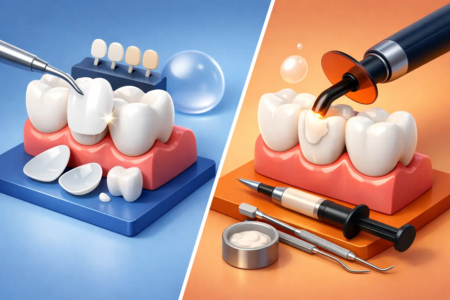
For implant dentistry, CBCT has become a major advantage. Placing an implant is not just about replacing a missing tooth. It is about positioning that implant in a way that supports function, appearance, and long-term stability.
A cone beam CT scan allows the dentist to measure bone height, width, and density with much greater accuracy. It also helps identify the location of nerves, sinus spaces, and neighboring roots. That level of detail supports precise implant placement, which can improve comfort, reduce surgical risk, and help create a more natural-looking result.
For patients considering full-arch solutions or same-day implant treatment, this planning becomes even more important. When multiple implants must work together to support a new smile, accuracy is everything.
Better insight for extractions and oral surgery
Not all extractions are straightforward. Wisdom teeth, severely damaged teeth, and teeth near nerves or sinus cavities can present challenges. A 3D scan helps your dentist or oral surgeon see root angulation, surrounding bone, and the position of nearby structures before treatment begins.
That can lead to a more efficient procedure and fewer unexpected findings during surgery. It can also help your dental team explain the treatment more clearly, which many patients appreciate, especially if they feel nervous about an upcoming procedure.
Stronger support for endodontic diagnosis
Root canal therapy depends on finding and treating the internal anatomy of the tooth as accurately as possible. Some teeth have extra canals, unusually narrow canals, or infections that are difficult to evaluate on standard X-rays.
CBCT can help identify hidden canals, root fractures, resorption, and the true extent of infection. In selected cases, that can make the difference between retreating a tooth successfully and missing the actual source of pain. It is not necessary for every root canal, but when symptoms are persistent or the anatomy is complicated, the added detail can be extremely helpful.
Clearer diagnosis can mean fewer surprises
Patients often want one simple answer before starting treatment: What exactly is wrong? One of the most practical benefits of cone beam CT dental imaging is that it can improve diagnostic confidence.
If a patient has ongoing pain with no obvious cause on a 2D X-ray, a cone beam scan may reveal a hidden infection, a vertical root fracture, impacted teeth, bone loss patterns, or sinus-related issues affecting the upper teeth. For orthodontic or airway-related concerns, it can also provide a broader picture of jaw relationships and skeletal structure.
That does not mean every patient needs this type of scan. It means that when the clinical situation is unclear, a more complete image can help avoid delayed diagnosis and reduce the chance of starting the wrong treatment.
Why 3D imaging can improve safety
Safety in dentistry often comes down to planning. The more accurately your dentist can locate nerves, roots, bone contours, and sinus spaces, the more carefully treatment can be tailored to your anatomy.
This is especially relevant in implant placement, surgical extractions, and other advanced procedures where millimeters matter. A CBCT scan helps minimize uncertainty and supports a more conservative, well-informed approach. For patients, that often translates into greater peace of mind.
There is also a comfort factor in simply knowing your provider is not estimating. In a practice that values precision and personalized care, advanced imaging helps turn a treatment plan into something far more specific to the individual sitting in the chair.
Are there trade-offs to consider?
Yes, and a thoughtful dental team should be transparent about them. Cone beam CT is not automatically the right choice for every appointment. Routine cleanings, basic cavity checks, and many standard evaluations can be handled very well with conventional digital X-rays and a clinical exam.
CBCT also involves radiation exposure, although the dose is generally much lower than a medical CT scan and varies depending on the machine, scan size, and purpose. The decision to use it should always be based on clinical need. In other words, the value comes from using the technology appropriately, not using it routinely just because it is available.
Cost can be another factor depending on the case and whether the scan is part of a larger treatment plan. For many patients pursuing implants, oral surgery, or complex restorative care, the added diagnostic value may justify that investment because it supports better outcomes and fewer complications. But the right choice depends on the procedure, the patient, and the level of detail required.
What patients can expect during a cone beam CT scan
The scan itself is fast and noninvasive. In most cases, you will remain standing or seated while the machine rotates around your head for a brief period. There are no needles, no recovery time, and no discomfort from the imaging process itself.
What matters most happens afterward. Your dentist reviews the scan carefully, evaluates the anatomy in three dimensions, and uses that information to guide diagnosis or planning. For many patients, this also makes the consultation easier to understand. When your dentist can show you the exact shape of the bone or the position of an impacted tooth, the conversation becomes more concrete and less abstract.
That clarity can be reassuring for patients who are weighing a major decision. Whether you are considering a single implant, a cosmetic-restorative plan, or more advanced rehabilitation, seeing the reason behind the recommendation often builds confidence.
When cone beam CT tends to be most useful
CBCT is commonly most valuable for dental implants, wisdom teeth assessment, complex extractions, root canal complications, TMJ evaluation, orthodontic planning, and full mouth rehabilitation. It can also be useful in cases involving trauma, unexplained pain, severe bone loss, or suspected pathology.
At a technology-forward practice such as San Clemente Dental Associates, this type of imaging supports the level of detail that complex and aesthetic cases often require. It fits especially well when treatment needs to balance health, function, and appearance, rather than addressing only one concern at a time.
The real point is not the machine itself. It is what the technology allows your dental team to do better: diagnose with more confidence, plan with more precision, and treat with a clearer understanding of your anatomy.
If you have been told you may need an implant, oral surgery, or another complex procedure, asking whether 3D imaging would improve the plan is a smart next step. The best dental care is not just about fixing a problem. It is about seeing the full picture before treatment begins.